Tissue Differentiation using IR Spectroscopy
The spectroscopic examination of organic material has commonly been conducted with FTIR spectrometers, but they are gradually being replaced by modern IR-spectrometers that are based on the principle of attenuated total reflection. Previous work has shown that this abundance of signals is not necessary to distinguish between malignant and benign tissue spectra. That is why only a few suitable quantum cascade lasers shall be combined and used in one sensor.
This yields several advantages:
- better signal-to-noise ratio
- lower recording time
- minimizing disruptive effects due to unwanted vibrations
The first goal in this project is to find the best suited wavelengths to achieve the best distinguishing result. Therefore, the following approaches are examined:
- analysis of IR spectra from literature, simulations, and experiments
- data analysis using multivariate statistical methods (e.g. principal component analysis)
- use of neural networks
Responsible: Felix Fischer
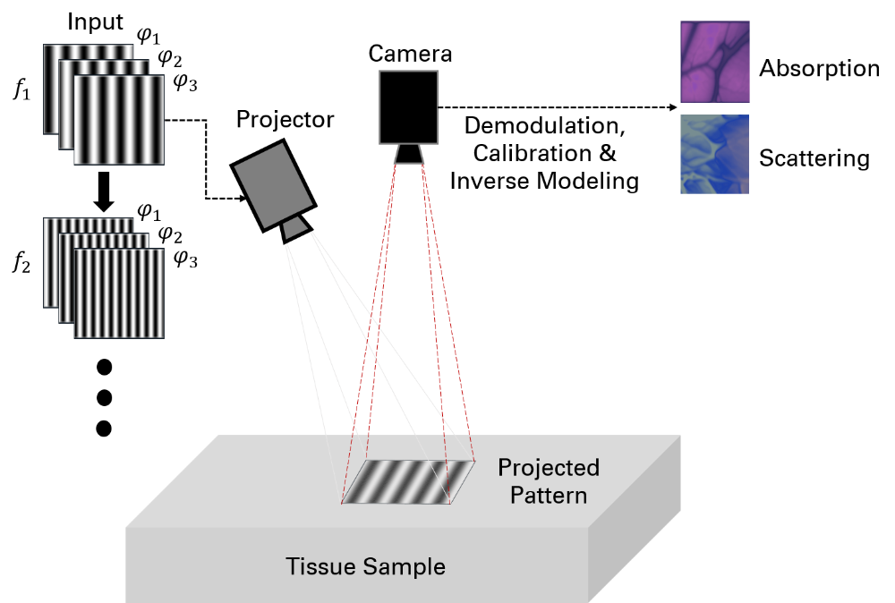
Fringe Projection Using 3D-Printed Micro-Optics for Model-Based Tissue Differentiation Based on Optical and Elastic Properties
The increased growth as well as the altered cell metabolism of tumors lead to a denser but less organized structure of the malignant tissue. The increased density of the extracellular matrix results in changes in the elastic parameters of the tissue composite. This serves as an approach to tissue differentiation since elasticity is quantifiable.
In addition, the scattering and absorption behavior of light is affected by irradiation into the tumor tissue. Biological tissue can be viewed as an optically diffuse, turbid medium, as light propagation within the tissue is usually dominated by scattering over absorption. Similarly to elastic parameters, these optical parameters can also be quantified. Based on chromophore concentrations in tissue, spatially resolved absorption and scattering coefficients can be determined metrologically. Due to altered cell metabolism and structure, malignant tissue contains different concentrations of these chromophores compared to healthy tissue.
The aim is the quantitative detection of the altered mechanical as well as the spatially resolved optical properties of the tissue by using model-based imaging techniques. The principle of fringe projection using 3D-printed micro-optics shall be miniaturized in such a way that both the elastic parameters due to deformation and the spatially resolved optical properties can be detected endoscopically.
Responsible: Ömer Atmaca
Contact

Ömer Atmaca
M.Sc.Research Assistant