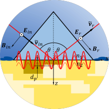
The spectroscopic examination of organic material has commonly been conducted with FTIR spectrometers, but they are gradually being replaced by modern IR-spectrometers that are based on the principle of attenuated total reflection. Previous work has shown that this abundance of signals is not necessary to distinguish between malignant and benign tissue spectra. That is why only a few suitable quantum cascade lasers shall be combined and used in one sensor.
This yields several advantages:
- better signal-to-noise ratio
- lower recording time
- minimizing disruptive effects due to unwanted vibrations
The first goal in this project is to find the best suited wavelengths to achieve the best distinguishing result. Therefore, the following approaches are examined:
- analysis of IR spectra from literature, simulations, and experiments
- data analysis using multivariate statistical methods (e.g. principal component analysis)
- use of neural networks
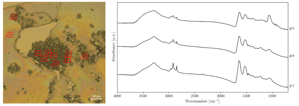
Microtome section of a bladder carcinoma organoid under an IR microscope (left). The red squares highlight the position and integration area of the IR spectroscopy. The spectra are displayed on the right.
Supported by: