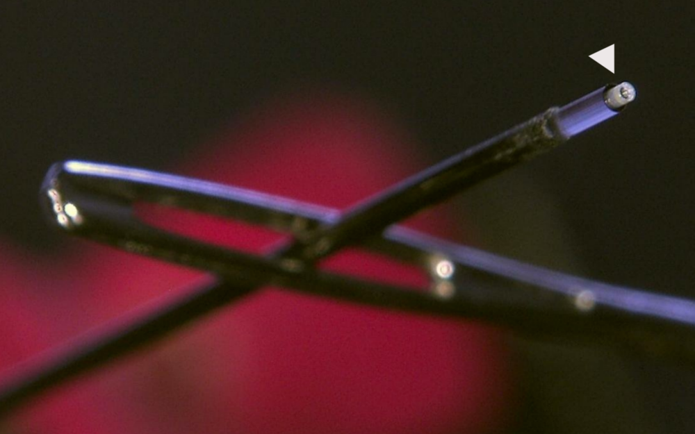
The smaller the diameter of an endoscope, the greater its potential for minimally invasive surgical treatments. In conventional, flexible (fiber-based) endoscopes with small diameters (<200 µm), imaging is severely limited by the number of fiber cores. Due to this limitation, the image is pixelated (image guide fiber) or, in the case of a single-mode fiber, consists of only a single pixel. In this project, an engineering approach is used to increase the number of pixels by spectral splitting, i.e. multiplying them. However, this requires very small color-splitting optical systems at the distal end of the endoscope, i.e., at the body-facing end of the fiber. Such small spectral optics systems are practically impossible to produce directly on the fiber using conventional techniques. In this project, therefore, the idea will be implemented using 3D-printed micro-optics. Preliminary work has shown that so-called multiphoton lithography (3D printing technique) is capable of producing imaging systems on this size scale. The aim of this project is thus to combine the bold idea of spectral pixel multiplication with the innovative manufacturing process of 3D printing, thus opening up new fields of application in medical technology with extremely small and also flexible endoscopes. In the medium term, this should enable improved diagnostics and treatment of strokes and heart attacks, for example (funded by the Vector Stiftung, Universität Stuttgart and the MWK).
Contact

Andrea Toulouse
Dr.Group leader 3D-printed Microoptics and Simulation